Brain UK study ref: 19/005,
Lay summary,
Project status: Closed
Spatial subtyping of glioblastoma using in situ sequencing (ISS)
Prof Mats Nilsson, Stockholm University, Sweden
Over the last 10 years it has been established that malignant tumours are not just large numbers of tumour cells that are similar to each other, but instead that there can be large differences between different regions and even between different groups of cells within the tumour. This has been called “tumour heterogeneity”, indicating that there are difference between individual tumour cells. Modern gene analysis methods can now find out how many different types of cells there are in the tumour and what type of genetic faults are within those tumour cells. In addition to point mutations (changes of single elements in the DNA), there are far more complex changes in the genes of cancer cells. The usual way of doing these tests is to mix tens of thousands of such tumour cells and do computer-based analysis. However, this will simply tell us how many cells with certain mutations are in the tumour. We will not learn how these are actually distributed, i.e. whether there are found across the entire tumour or a small islands only.
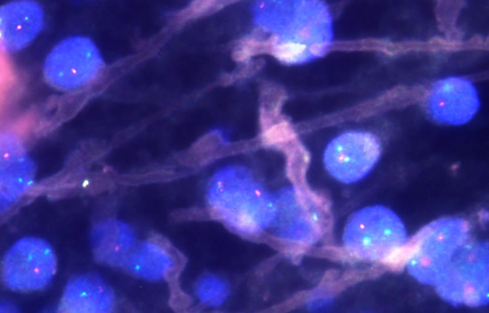
Our laboratory in Stockholm (Nilsson Group affiliated to Stockholm University at Stockholm’s Science for Life Laboratory) has developed a method which allows us to analyse exactly that: with colour-coded artificially engineered fragments of DNA (like a bar-coded fingerprint) we can literally see how cells with a certain mutation are distributed across the tumour. With this methodology we can for example look at a tumour from the first operation and see how a tumour after the second operation, for example after accumulating more mutations, has developed. We can also see how certain clones of tumour cells have disappeared or expanded. This will give us valuable information about therapy response in patients with gliomas.
