Brain UK study ref: 23/015,
Lay summary,
Project status: Active
Epidemiologic, Histologic, and Molecular Features of Methylation Class Pleomorphic Xanthoastrocytoma
Dr Kenneth Aldape and Dr Chris Dampier, National Institutes of Health, National Cancer Institute, USA
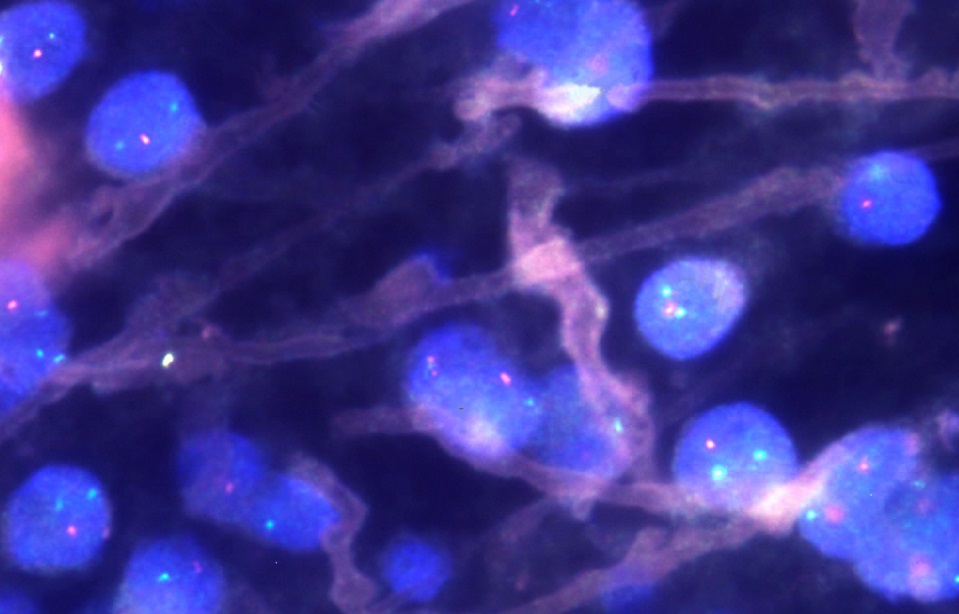
We want to make it easier for doctors to recognize a type of brain tumor called pleomorphic xanthoastrocytoma. The tumor gets its name because it is made of cells with a variety of shapes and sizes (pleomorphic) that sometimes contain fat droplets (xantho=yellow). Doctors used to recognize this tumor only by a set of patterns seen with a microscope. That was hard to do for two reasons. First, the patterns seen in this type of tumor are also seen in other types of tumors. Second, different doctors looking at the same patterns can recognize different tumor types. Therefore, a method for recognizing tumor types based on extra parts attached to DNA molecules was established. Now, doctors use computers to recognize this tumor and others by the patterns of extra parts on the tumor DNA molecules. Unfortunately, we can’t be sure the tumors recognized with the new method are the same as those recognized with the old method (that is, microscopes alone). We are conducting this study to figure out if the tumors are the same, how they may be different, and also to find better ways to recognize them.